| 货号: 28294 |
| 产品全名: CS1(DM9) 兔单克隆抗体 |
| 基因符号 SLAM7 (19A; CD319; CRACC; CS1) |
| 描述: CS1 antibody(DM9) 兔单克隆抗体 |
| 背景: Self-ligand receptor of the signaling lymphocytic activation molecule (SLAM) family. SLAM receptors triggered by homo- or heterotypic cell-cell interactions are modulating the activation and differentiation of a wide variety of immune cells and thus are involved in the regulation and interconnection of both innate and adaptive immune response. Activities are controlled by presence or absence of small cytoplasmic adapter proteins; SH2D1A:SAP and:or SH2D1B:EAT-2. Isoform 1 mediates NK cell activation through a SH2D1A-independent extracellular signal-regulated ERK-mediated pathway (PubMed:11698418). Positively regulates NK cell functions by a mechanism dependent on phosphorylated SH2D1B. Downstream signaling implicates PLCG1; PLCG2 and PI3K (PubMed:16339536). In addition to heterotypic NK cells-target cells interactions also homotypic interactions between NK cells may contribute to activation. However; in the absence of SH2D1B; inhibits NK cell function. Acts also inhibitory in T-cells (By similarity). May play a role in lymphocyte adhesion (PubMed:11802771). In LPS-activated monocytes negatively regulates production of proinflammatory cytokines (PubMed:23695528). |
| 经过测试的应用: ELISA; Flow Cyt |
| 推荐稀释比: Flow Cyt 1:100 |
| 种属反应性: Rabbit |
| 亚型: Rabbit IgG |
| 纯化: Purified from cell culture supernatant by affinity chromatography |
| 种属反应性: 人 CS1 |
| 成分: Lyophilized from sterile PBS, pH 7.4. 5 % – 8% trehalose is added as protectants before lyophilization. |
| 储存和运输: Store at -20°C to -80°C for 12 months in lyophilized form. After reconstitution, if not intended for use within a month, aliquot and store at -80°C (Avoid repeated freezing and thawing). |
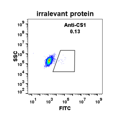 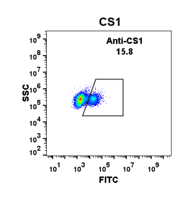
Figure 1. Expi 293 cell line transfected with irrelevant protein (left) and human CS1 (right) were surface stained with Rabbit anti-CS1monoclonal antibody 1µg/ml ( clone: DM9) followed by Alexa 488-conjugated anti-rabbit IgG secondary antibody. | 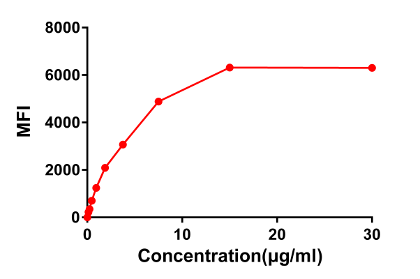
Figure 2. Flow cytometry data of serially titrated Rabbit anti-CS1 monoclonal antibody ( clone: DM9) on Raji cells. The Y-axis represents the mean fluorescence intensity (MFI) while the X-axis represents the concentration of IgG used. | 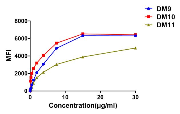
Figure 3. Affinity ranking of different Rabbit anti-CS1 mAb clones by titration of different concentration onto Raji cells. The Y-axis represents the mean fluorescence intensity (MFI) while the X-axis represents the concentration of IgG used. | 
Figure 4. Phylogenetic analysis of amino acid sequence of different Rabbit Anti-CS1 mAb clones. A) Heavy chain and B) Light chain. |
|