| 货号: 28361 |
| 产品全名: CD27(DM59) 兔单克隆抗体 |
| 基因符号 CD27; TNFRSF7; S152; T14; Tp55 |
| 描述: CD27 antibody(DM59) 兔单克隆抗体 |
| 背景: The protein encoded by this gene is a member of the TNF-receptor superfamily. This receptor is required for generation and long-term maintenance of T cell immunity. It binds to ligand CD70; and plays a key role in regulating B-cell activation and immunoglobulin synthesis. This receptor transduces signals that lead to the activation of NF-kappaB and MAPK8:JNK. Adaptor proteins TRAF2 and TRAF5 have been shown to mediate the signaling process of this receptor. CD27-binding protein (SIVA); a proapoptotic protein; can bind to this receptor and is thought to play an important role in the apoptosis induced by this receptor. |
| 经过测试的应用: ELISA; Flow Cyt |
| 推荐稀释比: ELISA 1:5000-10000; Flow Cyt 1:100 |
| 种属反应性: Rabbit |
| 亚型: Rabbit IgG |
| 纯化: Purified from cell culture supernatant by affinity chromatography |
| 种属反应性: 人 CD27 |
| 成分: Lyophilized from sterile PBS, pH 7.4. 5 % – 8% trehalose is added as protectants before lyophilization. |
| 储存和运输: Store at -20°C to -80°C for 12 months in lyophilized form. After reconstitution, if not intended for use within a month, aliquot and store at -80°C (Avoid repeated freezing and thawing). |
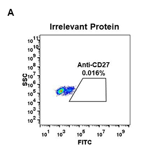 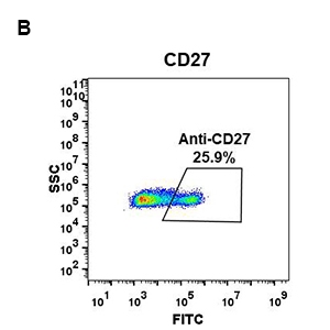
Figure 1. Expi 293 cell line transfected with irrelevant protein (A) and human CD27 (B) were surface stained with Rabbit anti-CD27 monoclonal antibody 1µg/ml ( clone: DM59) followed by Alexa 488-conjugated anti-rabbit IgG secondary antibody. | 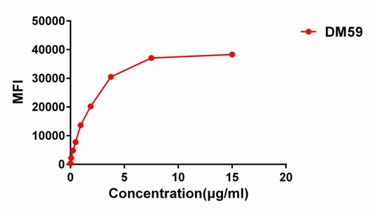
Figure 2. Flow cytometry data of serially titrated Rabbit anti-CD27 monoclonal antibody ( clone: DM59) on Raji cells. The Y-axis represents the mean fluorescence intensity (MFI) while the X-axis represents the concentration of IgG used. | 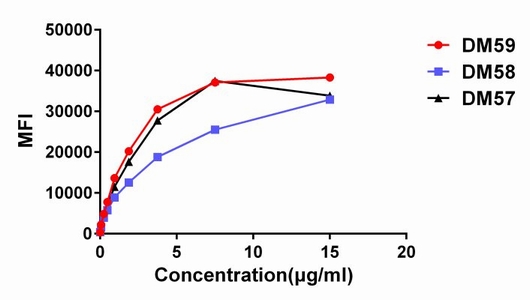
Figure 3. Affinity ranking of different Rabbit anti-CD27 mAb clones by titration of different concentration onto Raji cells. The Y-axis represents the mean fluorescence intensity (MFI) while the X-axis represents the concentration of IgG used. | |
|