| 货号: S217068 |
| 产品全名: IDH3G 兔多抗 |
| 基因符号 H-IDHG |
| UNIPROT ID: P51553 (Gene Accession – BC001902 ) |
| 背景: Isocitrate dehydrogenases catalyze the oxidative decarboxylation of isocitrate to 2-oxoglutarate. These enzymes belong to two distinct subclasses, one of which utilizes NAD(+) as the electron acceptor and the other NADP(+). Five isocitrate dehydrogenases have been reported: three NAD(+)-dependent isocitrate dehydrogenases, which localize to the mitochondrial matri,x and two NADP(+)-dependent isocitrate dehydrogenases, one of which is mitochondrial and the other predominantly cytosolic. NAD(+)-dependent isocitrate dehydrogenases catalyze the allosterically regulated rate-limiting step of the tricarboxylic acid cycle. Each isozyme is a heterotetramer that is composed of two alpha subunits, one beta subunit, and one gamma subunit. |
| 抗原: Fusion protein of human IDH3G |
| 经过测试的应用: ELISA, WB, IHC |
| 推荐稀释比: IHC: 25-100;WB: 500-2000;ELISA: 2000-5000 |
| 种属反应性: Rabbit |
| 克隆性: Rabbit Polyclonal |
| 亚型: Immunogen-specific rabbit IgG |
| 纯化: Antigen affinity purification |
| 种属反应性: Human, Mouse, Rat |
| 成分: PBS (without Mg2+ and Ca2+), pH 7.4, 150 mM NaCl, 0.05% Sodium Azide and 40% glycerol |
| 研究领域: Metabolism |
| 储存和运输: Store at -20°C. Avoid repeated freezing and thawing |
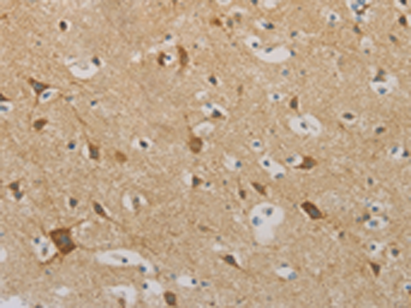
Immunohistochemistry analysis of paraffin embedded Human brain tissue using 217068(IDH3G Antibody) at a dilution of 1/30(Cytoplasm). | 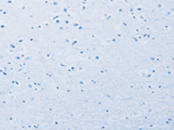
In comparision with the IHC on the left, the same paraffin-embedded Human brain tissue is first treated with the fusion protein and then with 217068(Anti-IDH3G Antibody) at dilution 1/30. | 
The image on the left is immunohistochemistry of paraffin-embedded Human thyroid cancer tissue using 217068(Anti-IDH3G Antibody) at a dilution of 1/30. | 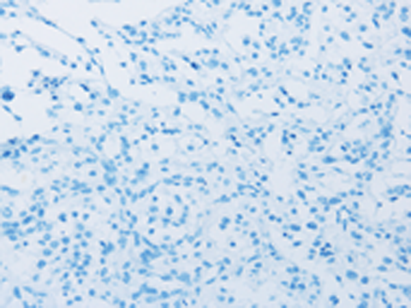
In comparision with the IHC on the left, the same paraffin-embedded Human thyroid cancer tissue is first treated with fusion protein and then with D221734(Anti-IDH3G Antibody) at dilution 1/30. | 
Gel: 10%SDS-PAGE, Lysate: 40 μg;
Lane 1-2: Mouse brain tissue, NIH/3T3 cells;
Primary antibody: 217068(IDH3G Antibody) at dilution 1/350;
Secondary antibody: Goat anti rabbit IgG at 1/8000 dilution;
Exposure time: 15 seconds | |
|